
 (585) 768-2513
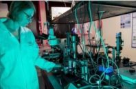
A three-year collaboration involving researchers at the University of Nottingham (Nottingham, England; the collaboration leader), Heriot-Watt University (Edinburgh, Scotland), and the University of Glasgow (Glasgow, Scotland) will develop an instrument that combines four optical microscopy technologies to image live cancer cells in 4D. The multimodal device will also plot, track, and analyze the cells’ position, movement, and function as they grow into and interact with the proteins and sugars that make up their surrounding environment (the extracellular matrix).
This will enable scientists to understand how physical and biochemical cues from cells influence the matrix, and vice versa, and how these interactions affect the progression of disease.
“While a conventional microscope produces a sharp image from just a single object-plane, multiplaning records sharp images from several object planes simultaneously,” says Paul Dalgarno of Heriot-Watt University, who pioneered a multiplane imaging technique that underpins the new instrument. “We will combine our revolutionary multiplane system with optical trapping before integrating this into the full light sheet-based trapping system of our co-collaborators. Together, this will enable us to fully study how cells migrate, grow, and interact in 3D and in real time.”
