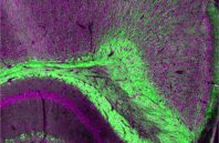
To a patient, the analysis of a tissue biopsy sample to check for something like cancer may seem like a relatively simple process, even if it does mean giving up a small piece of flesh to be tested. The sample heads off to a lab, the patient heads home, and in several days the doctor calls with the results.
In reality, quite a bit of work goes into preparing a tissue sample and evaluating it for signs of disease. To be viewed under a microscope, the sample needs to be cut into extremely thin slices that might be only a few cells thick. And to aid with the viewing, the technician may employ a variety of dyes to label specific proteins or cell structures.
“Extensive processing of the sample is required,” says Lihong Wang, Caltech’s Bren Professor of Medical Engineering and Electrical Engineering in the Division of Engineering and Applied Science. “You can only label so many molecules at a time, and you have to do a washing between labelings. And some molecules don’t absorb dyes and don’t get labeled at all.”

 (585) 768-2513

