
 (585) 768-2513
A Virginia Commonwealth University researcher has developed a procedure for identifying the source of cells present in a forensic biological sample that could change how cell types are identified in samples across numerous industries.
Many traditional techniques for distinguishing between saliva, blood, skin or vaginal tissue in an evidence sample are based on microchemical reactions that can be prone to false-positive or false-negative results, according to the researcher, Christopher Ehrhardt, Ph.D., an associate professor in the Department of Forensic Science in the College of Humanities and Sciences. Additionally, they may be difficult to use on aged or heavily degraded samples.
“The information is often limited,” Ehrhardt said. “And when using conventional methods, you have to be prepared to consume part of the sample in most cases, which decreases the value of it.”
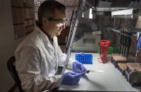
Ehrhardt’s procedure aims to change that. He begins by taking microscopic images of the individual cells using a benchtop microscope or a flow cytometer—a device used in cell biology that photographs individual cells encased within drops of water. Ehrhardt then makes measurements that capture size, shape and fluorescent properties of the cells. Those measurements are then analyzed using machine learning algorithms—in this case computer software programmed to recognize characteristics of the images—to correlate them with cell type.
