
 (585) 768-2513
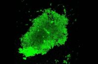
A team of researchers at the Stanford University School of Medicine (Stanford, CA) has developed a two-piece fluorescent probe that is activated when it comes in contact with tuberculosis (TB) bacteria in phlegm. The technique can diagnose live TB in an hour and help monitor the efficacy of treatments.
Jianghong Rao, Ph.D., a professor of radiology at the School of Medicine and the senior author of a paper describing the work, says that current methods for TB diagnosis can take up to two months to complete—a stretch during which infected individuals could spread the disease broadly, even if they don’t know they are infected. A quicker diagnosis could curtail the infection rate. What’s more, the new method is cheaper and easier to carry out, ideally enabling healthcare providers in poorer communities to one day adopt the technology.
To diagnose TB, clinicians need to collect a sample of sputum, cultivate it in the lab, and wait for the bacteria to grow to detectable level. It also requires specialized facilities, which are missing in many hospitals worldwide. The new imaging technique, however, uses conventional fluorescence microscopes that nearly all hospitals have and that require no special training, Rao says. All that is needed is a sample of the patient’s sputum that can be prepared and put under the microscope for analysis.
