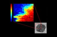
A team at the University of Lyon has developed a light-scattering method that maps out the mechanical properties of a tumor’s cellular structure as well as its internal fluids, revealing changes due to chemotherapy treatment. The technique could be used to differentiate populations of malignant cells and monitor how effective an anticancer treatment is.
The research team used a noncontact imaging technique that exploits the minute vibrations of matter that occur naturally. The team’s technique does not require the use of contrast agents and therefore does not disturb tissue function.
To replicate the behavior of colorectal tumors in vitro, the researchers created organoids made from tumor cells. They focused a red laser beam onto the organoids. They found that infinitesimal vibrations of these samples, generated by thermal agitation, slightly affected the color of the light beam as it passed through and exited the sample. By analyzing this light, the team was able to construct images showing the variations in mechanical properties within the tumor/organoid.

 (585) 768-2513

