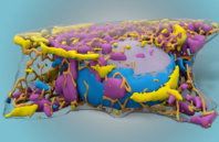
Scientists at the Allen Institute have used machine learning to develop a label-free tool for predicting 3D fluorescence directly from transmitted-light images. Instead of fluorescence microscopy, the new method uses only black-and-white images generated by a bright-field microscope.
To the human eye, cells viewed in a bright-field microscope are sacs rendered in shades of gray. A trained scientist can find the edges of a cell and the nucleus, but not much else. Using a convolutional neural network, the team trained computers to recognize the details in the bright-field images. The researchers tested 12 different cellular structures and were able to show that their technique could be used to generate multistructured, integrated images. The model generated by the machine learning algorithm predicted images that matched the fluorescently labeled images for most of the structures.

 (585) 768-2513

