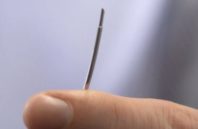
A team of researchers at University College London (UCL) and Queen Mary University of London (QMUL) has developed a new optical ultrasound needle that can image heart tissue in real time during keyhole procedures. The technology has been used for minimally invasive heart surgery in pigs, giving a high-resolution view of soft tissues up to 2.5 cm in front of the instrument, inside the body.
Doctors currently rely on external ultrasound probes combined with preoperative imaging scans to visualize soft tissue and organs during keyhole procedures, as the miniature surgical instruments used do not support internal ultrasound imaging. Recognizing this limitation, the research team designed and built the optical ultrasound technology to fit into existing single-use medical devices, such as a needle.

 (585) 768-2513

