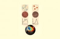
A team of scientists from Helmholtz Zentrum München, the Jülich Research Center, the Technical University of Munich, and Heinrich Heine University Düsseldorf (all in Germany) has shown that harmless purple bacteria of the genus Rhodobacter are capable of visualizing aspects of tumor heterogeneity in cancerous tumors. With the aid of optoacoustic imaging, the researchers used these microorganisms to visualize cells of the immune system known as macrophages, which also play a role in tumor development.
Many cancers form solid tumors that reveal major differences at the cellular and molecular level. One of these concerns the localization and activity of macrophages. Although these cells are essential for a healthy immune system, they also play a key role in tumor development. With the aid of photosynthetic bacteria, new optoacoustic techniques, which indicate where such macrophages are present and active, have now been developed.
“We were able to demonstrate that bacteria of the genus Rhodobacter, which are harmless to humans, are suitable as indirect markers of macrophage presence and activity,” says Dr. Andre C. Stiel, head of the Cell Engineering Group at the Institute of Biological and Medical Imaging (IBMI) at Helmholtz Zentrum München. Rhodobacter bacteria produce large quantities of the photosynthetic pigment bacteriochlorophyll a. This pigment enabled the researchers to detect bacteria in a tumor by means of multispectral optoacoustic tomography (MSOT).

 (585) 768-2513

