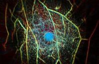
Knowing that the conventional method for breast cancer screening—mammography—is less than ideal for multiple reasons (including exposure to x-ray radiation and discomfort for patients), researchers at the California Institute of Technology (Caltech; Pasadena, CA) have developed a laser-sonic scanner that can find tumors in as little as 15 seconds by shining pulses of light into the breast.
The scanning system, known as photoacoustic computed tomography (PACT), works by shining a near-infrared laser pulse into the breast tissue. The laser light diffuses through the breast and is absorbed by oxygen-carrying hemoglobin molecules in the patient’s red blood cells, causing the molecules to vibrate ultrasonically. Those vibrations travel through the tissue and are picked up by an array of 512 ultrasonic sensors around the skin of the breast. The data from those sensors are used to assemble an image of the breast’s internal structures in a process that is similar to ultrasound imaging, though much more precise. PACT can provide a clear view of structures as small as a quarter of a millimeter at a depth of 4 cm. Mammograms cannot provide soft-tissue contrast with the level of detail in PACT images, explains Lihong Wang, Caltech’s Bren Professor of Medical Engineering and Electrical Engineering, whose lab developed the technique.

 (585) 768-2513

