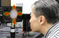
Fundus photography, a standard imaging tool used by ophthalmologists, has existed for almost two centuries. However, its use in remote and low-resource regions, where traveling to a clinic is not always practical, has been limited.
A compact eye fundus camera system has been developed that allows a user to photograph retinal images of the interior of the eye by using high-speed image-processing NIR light. At 2.3 mm2, the system is small enough to fit on a smartphone. It can be mounted on a smartphone without compromising the power necessary for capturing highly detailed images of the interior lining of the eye.
Created by researchers at Nara Institute of Science and Technology (NAIST), the camera can achieve 1,000 images per second. This ensures that the camera can quickly align itself with constant changes in the path of the light that travels through the retina to the back of the eye.
To ensure that the light from the camera is strong enough to illuminate the interior of the eye, researchers used a modified CMOS sensor. The miniaturized sensor incorporates three NIR filters, which acquire three signals that can be given a red, green, or blue value, to generate a color photograph of the eye.

 (585) 768-2513


Very interesting – I work in the biophotonics field especially in the area of eye safety and I believe that fundus inspection could be very important for everyone in the near future, especially as we worry about macula degeneration and so on.
Very good luck with this project.
Best regards
Dr Neil Haigh
Blueside Photonics Ltd