
 (585) 768-2513
Several bioimaging techniques are potentially able to image the metabolic process underway in living cells, but many of them have some inherent drawbacks, either in terms of their limited spatial resolution or the need to introduce potentially toxic labeling molecules.
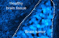
A team led by Wei Min at Columbia University has now developed a promising new alternative, by combining stimulated Raman scattering with the use of relatively innocuous deuterium oxide—or heavy water—as a contrast agent, one which is readily incorporated into proteins and lipids during metabolic activities.
The new method, termed deuterium oxide probing and stimulated Raman scattering (DO-SRS), should offer high sensitivity and subcellular resolution, while also being suitable for in vivo live imaging in mammals. The work was published in Nature Communications.
