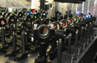
To better understand how proteins move and interact within a cell membrane, an imaging method called live-cell single fluorescent-molecule imaging (SFMI) can be used to tag each protein in the membrane with a fluorescent marker. The proteins are visible under a custom-built fluorescence microscope. However, the markers under the microscope photobleach over time; because of this, researchers using SFMI have only been able to observe individual molecules for about 10 s at a time.
Researchers at Okinawa Institute of Science and Technology (OIST) have developed a way to suppress photobleaching when using SFMI. Researchers placed the cells in an environment that mimicked the actual conditions inside a living organism. To this environment, which had low concentrations of dissolved oxygen, they added the antioxidants trolox and trolox quinone. This reducing-plus-oxidizing system, combined with low oxygen concentrations, was able to strongly suppress photobleaching, with only minor effects on the cells.

 (585) 768-2513

