
 (585) 768-2513
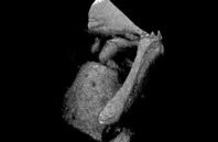
A research team led by OSA Fellow Seok-Hyun Yun at the Wellman Center for Photomedicine at Massachusetts General Hospital, USA, reports using a form of optical coherence tomography (OCT) to capture 3-D anatomical and vibrational images of ossicles—tiny bones within the middle ear—as they move in response to sound. The imaging method, which they named OCT vibrography, uses OCT phase-synchronized with a range of high-frequency sound stimuli. In demonstrations with a chinchilla ear model, the researchers used OCT vibrography to observe a previously unknown mode of ossicular motion at high frequencies.
The new instrument, which is an improvement on the team’s previous design, may offer insights into how mechanical vibrations of ossicles contribute to sound perception in the brain. According to the team, it could someday also be used to diagnose hearing problems.
